Pupilla learning lab · Anatomy
3D Eye Anatomy Simulator
Pupilla’s 3D eye anatomy simulator lets you rotate, slice, separate and label a single eye model — from the tear film and the ten retinal layers to the six extraocular muscles and the bony orbit — and move it through gaze, pupil and accommodation states.
- Free · no install
- English & Arabic
- Works on phone and desktop
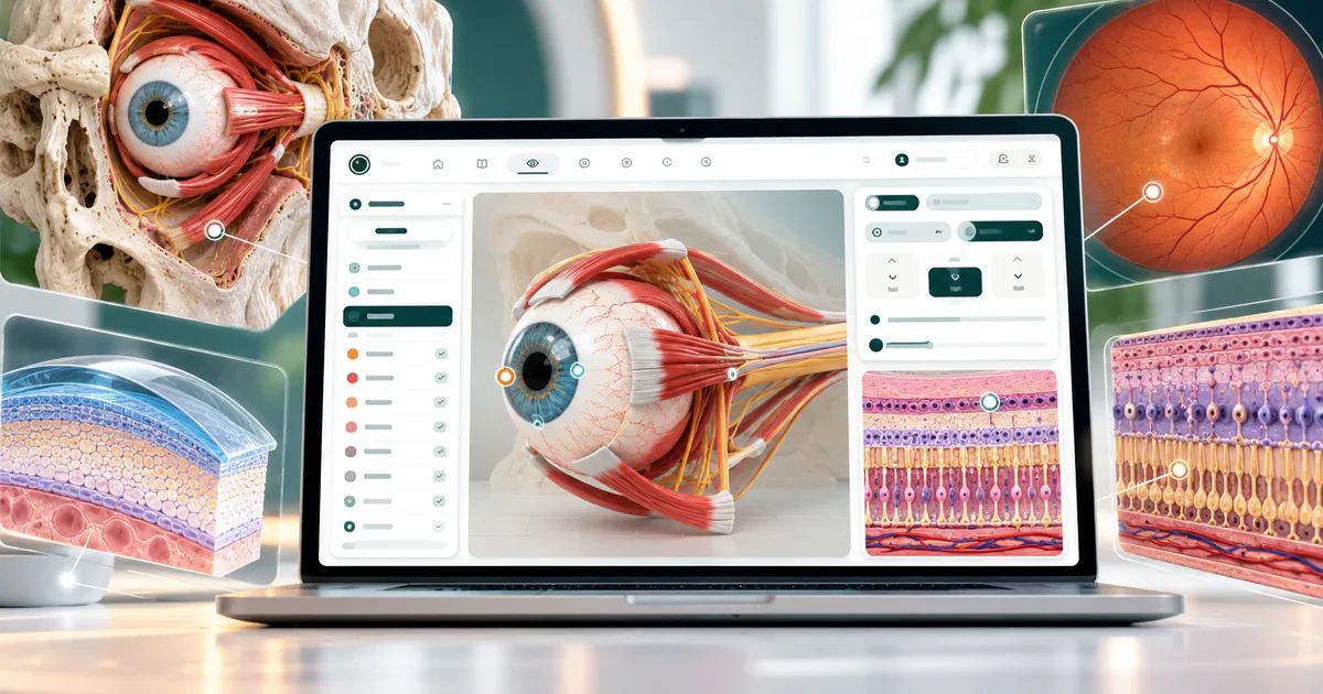
The 3D eye model at a glance
| Feature | In the model |
|---|---|
| Selectable structures | 42 — coats, anterior segment, lens, ciliary region, fluids, retinal circulation, muscles and optic nerve |
| Study modes | 8 — whole eye, dissection, anterior segment, fundus, retinal layers, foveal section, cornea study, orbit study |
| Retinal layers | 10 retinal layers (ILM to RPE) plus Bruch’s membrane and choroid as adjacent tissue |
| Corneal layers | 6 — tear film, epithelium, Bowman’s layer, stroma, Descemet’s membrane, endothelium |
| Extraocular muscles | 6 per eye, with tendons, the trochlea and the common tendinous ring |
| Orbit | 7 orbital bones, 19 study presets, 6 camera views, intraconal and extraconal fat |
| Live measurements | Pupil 2–8 mm, accommodation, corneal thickness and white-to-white, axial and diameter overlays |
| Languages & price | English and Arabic (RTL) · free · no install |
Eight ways to study the eye
- 01 · Whole eyeRotate and zoom the complete globe with its muscles, optic nerve and vessels. Select any part to read its name and function, or search a structure by name.
- 02 · DissectionCut the eye on sagittal, coronal, horizontal, internal or cutaway planes with closed section faces, then slide the “separate structures” control to pull the layers apart.
- 03 · Anterior segmentCornea, limbus, iris, pupil, lens and capsule, zonules, ciliary body, and the drainage angle: Schwalbe’s line, trabecular meshwork, scleral spur and Schlemm’s canal.
- 04 · FundusLook from inside the globe at the optic disc, macula, fovea and the retinal arterial and venous arcades.
- 05 · Retinal layersPeel the retina layer by layer, from the internal limiting membrane to the pigment epithelium, with cell and fibre patterns for the nuclear and plexiform layers.
- 06 · Foveal sectionA cross-section through the foveal pit where the inner layers part and the outer nuclear layer and photoreceptor segments thicken.
- 07 · Cornea studySix selectable layers with true physical thickness, cut sections, layer separation, a 0.9 mm central microscopic crop, curvature and pachymetry maps, and a reversible keratoconus teaching example.
- 08 · Orbit studyThe skull and both orbits, the seven orbital bones, the muscle cone, orbital apex, and intraconal and extraconal fat, with muscle attachments.
All 42 selectable structures
Every structure can be selected, isolated, hidden, made transparent, labelled and searched by name. Grouping follows the model’s own anatomy panel.
| Group | Structures |
|---|---|
| Ocular coats | Sclera · choroid · retinal pigment epithelium · retina |
| Anterior segment | Cornea · limbus · iris · pupil aperture · sphincter pupillae · dilator pupillae · Schwalbe’s line · trabecular meshwork · scleral spur · Schlemm’s canal |
| Lens & suspension | Crystalline lens · lens capsule · zonular fibres |
| Ciliary region | Ciliary body · ciliary muscle · ciliary processes · pars plana |
| Posterior landmarks | Macula · fovea centralis · optic disc |
| Chambers & vitreous | Anterior chamber · posterior chamber · vitreous body |
| Retinal circulation | Retinal arteries and veins · central retinal artery · central retinal vein · episcleral vessels |
| Motor system | Superior, inferior, medial and lateral recti · superior and inferior obliques |
| Optic nerve & support | Optic nerve · optic nerve sheath · lamina cribrosa · trochlea and common tendinous ring |
Retinal layers in 3D
Ten retinal layers from the vitreous outward, drawn with a foveal pit where the inner layers part. Bruch’s membrane and the choroid are shown as neighbouring tissue, not as retinal layers.
| Layer | Name | What it is |
|---|---|---|
| ILM | Internal limiting membrane | Vitreoretinal boundary associated with Müller cell endfeet. |
| NFL | Nerve fibre layer | Ganglion cell axons running toward the optic disc. |
| GCL | Ganglion cell layer | Ganglion cell bodies; their axons form the NFL. |
| IPL | Inner plexiform layer | Synapses among bipolar, amacrine and ganglion cells. |
| INL | Inner nuclear layer | Nuclei of bipolar, horizontal, amacrine and Müller cells. |
| OPL | Outer plexiform layer | Photoreceptor synapses; oblique cone axons form Henle’s fibre layer at the macula. |
| ONL | Outer nuclear layer | Rod and cone nuclei; densely packed cone nuclei at the foveal centre. |
| ELM | External limiting membrane | A junctional belt between Müller cells and photoreceptors, not a free sheet. |
| PR | Photoreceptor segments | Inner and outer segments of rods and cones facing the RPE; the foveal centre is rod-free. |
| RPE | Retinal pigment epithelium | Supports photoreceptors and forms the outer blood–retinal barrier. |
| BrM | Bruch’s membrane (adjacent) | Interface between the RPE and choriocapillaris; outside the ten retinal layers. |
| CH | Choroid (adjacent) | Vascular coat external to Bruch’s membrane that supplies the outer retina. |
Corneal layers and the cornea study
The cornea study samples one shared surface model, so layers, thickness, curvature and maps always agree. Thicknesses below are the model’s true physical values.
| Layer | Thickness | Notes |
|---|---|---|
| Tear film | ≈ 3 µm | Outside the central corneal thickness measurement. |
| Epithelium | ≈ 55 µm | Stratified: superficial squamous, wing and basal cells. |
| Bowman’s layer | ≈ 18 µm | Acellular layer beneath the epithelium. |
| Stroma | remainder | The bulk of corneal thickness: the remainder after the thin layers. |
| Descemet’s membrane | ≈ 10 µm | Basement membrane of the endothelium; its end marks Schwalbe’s line. |
| Endothelium | ≈ 4.5 µm | Single cell layer facing the anterior chamber. |
- Curvature, pachymetry and posterior-elevation teaching maps
- Cut sections with closed faces and layer separation
- A 0.9 mm central microscopic crop
- Central thickness, white-to-white and principal meridians you can change
- A reversible keratoconus teaching example and a side-by-side comparison with the reference cornea
Extraocular muscles: nerves, insertions and actions
Insertion distances from the limbus are typical adult values used by the model (the spiral of Tillaux). Primary action describes the muscle in primary gaze; several actions change with gaze direction, which you can test in the Motion laboratory.
| Muscle | Cranial nerve | Insertion from limbus | Primary action | Secondary actions |
|---|---|---|---|---|
| Medial rectus (MR) | III | 5.5 mm | Adduction | None significant |
| Lateral rectus (LR) | VI | 6.9 mm | Abduction | None significant |
| Superior rectus (SR) | III | 7.7 mm | Elevation (best in abduction) | Intorsion, adduction |
| Inferior rectus (IR) | III | 6.5 mm | Depression (best in abduction) | Extorsion, adduction |
| Superior oblique (SO) | IV | via the trochlea | Intorsion | Depression (best in adduction), abduction |
| Inferior oblique (IO) | III | from the anterior orbital floor | Extorsion | Elevation (best in adduction), abduction |
Motion laboratory
Set the eye to primary gaze, elevation, depression, adduction, abduction or diagonal gaze, adjust torsion, change the pupil from 2 to 8 mm, and trigger the near response. Each state names the responsible muscle and nerve and gives a clinical cue.
Study these actions in the extraocular muscles guide, then see how both eyes coordinate in the 3D eye movement simulator and test them clinically in the H-test guide.
The orbit: seven bones, muscle cone and fat
The orbit study has 19 presets, six camera views and six layer toggles (bones, globe, muscles, optic nerve, intraconal fat, extraconal fat), including an exploded view of the bones.
| Bone | What it contributes |
|---|---|
| Frontal | Most of the orbital roof and superior rim; the trochlea lies on its anteromedial surface. |
| Sphenoid | Posterior roof and lateral wall, around the apex and superior orbital fissure. |
| Zygomatic | Anterior lateral wall and part of the floor and rim. |
| Maxilla | Most of the floor; the inferior oblique arises just lateral to the lacrimal groove. |
| Palatine | Small orbital process in the posterior floor near the apex. |
| Lacrimal | Thin anterior medial-wall bone beside the lacrimal sac fossa. |
| Ethmoid | Lamina papyracea forms much of the medial wall, with the ethmoid air cells medial to it. |
Biometry and physiology
Explore the eye’s dimensions with diameter and axial-distance overlays, set the pupil between 2 and 8 mm, change accommodation, and separate the cornea and limbus, the lens and capsule, or the retina for inspection.
A six-step study route
- Start in Whole eye: rotate the model, then use the Globe, Muscles, Nerves and Vessels toggles to isolate one system.
- Switch to Dissection, choose a section plane and drag “separate structures” to see how the coats sit on one another.
- Open Retinal layers and name the ten layers from the vitreous outward; then open the Foveal section and explain which layers are displaced.
- Use the Cornea study to compare layer thicknesses, then change corneal thickness or curvature in Biometry and watch the maps respond.
- In the Motion laboratory, move the eye through the gaze positions and name the muscle responsible before reading the answer.
- Finish in the Orbit study: identify the seven bones, the muscle cone and where each fat compartment lies.
Who it is for
- Optometry and ophthalmology students learning gross anatomy and ocular histology
- Medical students and residents revising for anatomy, neuro-ophthalmology and strabismus
- Orthoptists and technicians who need the muscles, nerves and gaze positions in one model
- Educators who want a shared, language-switchable 3D reference for lectures and tutorials
Accuracy, sources and limits
Trust depends on knowing what a model does not claim.
- The model is a reference adult educational model, not a scan of a patient or a population average; it carries no clinical validation claim.
- Some elements are schematic by design: retinal layer thickness is in display units, vessel trees are representative, trabecular pores are enlarged, and display separation distances are not anatomical displacements.
- The optic-canal view in the orbit study is marked registration pending; it is shown as unavailable rather than guessed.
- Biometry and cornea readouts are calculated from the model for teaching. They are not device measurements and must not be used for IOL calculation, surgery or diagnosis.
For education and training only. Pupilla is not a medical device and must not be used for diagnosis or treatment decisions.
Frequently asked questions about the 3D eye model
What is the Pupilla 3D eye anatomy simulator?
A free, browser-based interactive 3D model of the human eye for education. You can rotate, zoom, slice and label 42 structures, study the retina and cornea layer by layer, move the eye through gaze positions, and explore the orbit and its bones.
How many layers of the retina does the model show?
Ten retinal layers, from the internal limiting membrane to the retinal pigment epithelium, plus Bruch’s membrane and the choroid shown as adjacent tissue. A foveal cross-section shows how the inner layers are displaced at the pit.
What are the layers of the cornea in the 3D model?
Six selectable layers: tear film, epithelium, Bowman’s layer, stroma, Descemet’s membrane and endothelium, drawn at true physical thickness by default, with an optional enhanced view that is clearly labelled.
Does it show the extraocular muscles and their actions?
Yes. All six muscles are modelled with tendons, the trochlea of the superior oblique and the common tendinous ring. The Motion laboratory moves the eye through elevation, depression, adduction, abduction, diagonal gaze and torsion so you can see which muscle acts.
Which nerve supplies each extraocular muscle?
Lateral rectus: cranial nerve VI. Superior oblique: cranial nerve IV. Medial, superior and inferior recti and the inferior oblique: cranial nerve III — remembered as LR6 SO4 R3.
Which bones form the orbit?
Seven: frontal, sphenoid, zygomatic, maxilla, palatine, lacrimal and ethmoid. The orbit study separates them, adds the globe, muscles and optic nerve, and shows intraconal and extraconal fat.
Can I see the fovea and macula in 3D?
Yes. The fundus mode looks from inside the globe at the optic disc, macula and fovea with the retinal vessel arcades, and the foveal section cuts through the pit to show its layer arrangement.
Does it show the aqueous outflow pathway?
The anterior segment includes the drainage angle: Schwalbe’s line, trabecular meshwork, scleral spur and Schlemm’s canal, along with the ciliary processes that secrete aqueous humour.
Is the pupil and accommodation interactive?
Yes. Pupil diameter can be set between 2 and 8 mm, miosis and mydriasis are shown with the sphincter and dilator pupillae, and near accommodation shows the lens becoming more convex as the ciliary muscle contracts. Accommodation is qualitative.
How accurate is the model?
It is built from published anatomical references and typical adult dimensions, with a shared measurement engine so cornea, biometry and layers stay consistent. It is an educational reference adult, not patient-specific, and parts that are schematic are labelled as such.
Does it work on a phone, and is there an Arabic version?
It runs in modern phone, tablet and desktop browsers; a larger screen helps for the orbit and cornea studies. The interface and structure descriptions are available in English and Arabic, with right-to-left layout.
Is it free, and do I need an account?
It is free and needs no installation or sign-in to explore the 3D anatomy.
Can it be used for diagnosis or surgical planning?
No. It is for education and training only and is not a medical device.
Related guides
Extraocular muscles: anatomy, actions and nerve supply
Six extraocular muscles move each eye: the medial, lateral, superior and inferior recti, and the superior and inferior obliques. The medial and lateral recti move the eye horizontally; the superior and inferior recti mainly elevate and depress it; the obliques mainly rotate it (torsion) and also move it vertically. The lateral rectus is supplied by the sixth cranial nerve, the superior oblique by the fourth, and all the others by the third — remembered as "LR6 SO4 R3".
Ocular motility and the H-test: ductions, versions and the nine positions of gaze
The H-test examines eye movements by asking the patient to follow a target in an H shape through the six cardinal positions of gaze. In each of those positions one muscle of each eye is the main mover, so an eye that lags in a position points to weakness of the muscle that works best there. Versions are movements of both eyes together, ductions are movements of one eye with the other covered, and vergences move the eyes in opposite directions. Adding straight up, straight down and primary position gives the nine positions of gaze, which also reveal A and V patterns.
Third, fourth and sixth cranial nerve palsies and the three-step test
A third nerve palsy causes ptosis and an eye that rests down and out, with limited elevation, depression and adduction, and sometimes a dilated pupil. A fourth nerve palsy weakens the superior oblique, causing a hypertropia of the affected eye that increases on gaze to the opposite side and on head tilt toward the same side, often with a compensatory head tilt away from the affected side. A sixth nerve palsy weakens the lateral rectus, causing an esotropia and limited abduction that increase on gaze toward the affected side and at distance.
Visual pathway lesions and the visual field defects they cause
The pattern of a visual field defect shows where the visual pathway is damaged. A lesion in front of the optic chiasm affects one eye; a lesion of the chiasm typically causes a bitemporal hemianopia; and any lesion behind the chiasm causes a homonymous defect on the opposite side in both eyes. The further back the lesion, the more congruous — alike in the two eyes — the defect becomes, so an occipital lesion gives a congruous homonymous hemianopia, often with macular sparing.
References & further learning
Sources for the teaching principles. Listing a source does not imply its institution endorses or validates this simulator.